Most clinicians believe that Keratoconus is inherited. It most often starts in the early teens. For this reason, parents who have the condition are encouraged to start checking their children for the disease at age 10.
What is Keratoconus?
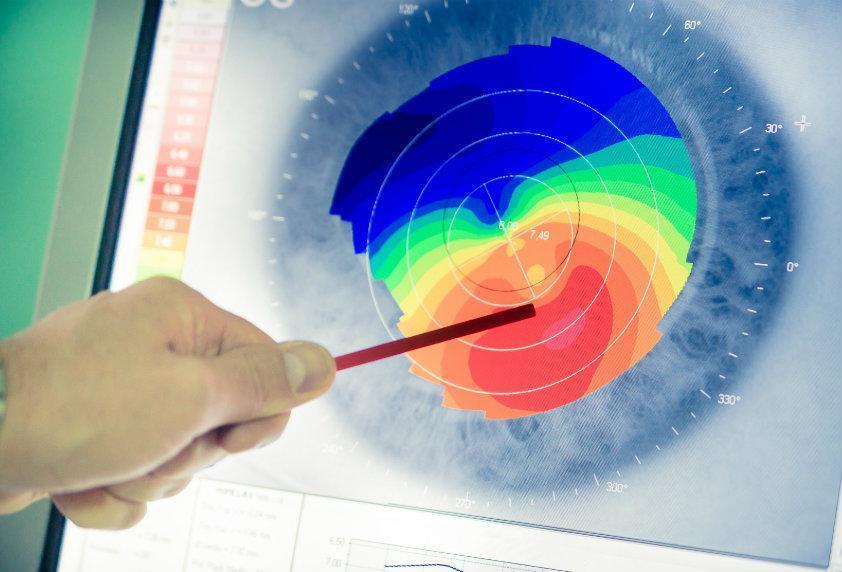
Keratoconus causes harmful changes to the shape of the cornea over a period of time. These changes can come on rapidly, or they may take several years to develop. The changes may also stop at any time, or they can go on changing the shape of the cornea for decades.
In severe cases, stretched fibers of collagen can cause terrible scarring. If the scarring is sufficiently pronounced, the back of the cornea can tear. This will cause swelling that can take months to subside.
Contributing conditions and risk factors
- Forcefully rubbing the eyes, (a common problem among children with allergies)
- Other eye conditions such as vernal keratoconjunctivitis, retinitis pigmentosa, and retinopathy of prematurity
- Other disorders such as enzyme abnormalities or heritable factors like Down’s syndrome
- Prolonged use of contact lenses
- Certain disorders such as Leber’s congenital amaurosis, Down’s syndrome, osteogenesis imperfect, and Ehlers-Danlos syndrome
Treatments
Treatment for Keratoconus usually begins with a new prescription for eyeglasses. If wearing these specially prescribed glasses does not provide serviceable vision improvements, contact lenses may be used instead. Rigid contact lenses that are gas permeable are usually the best for those with this condition.
In milder cases, new glasses will usually be enough to restore clear vision to the patient. In time, however, it will most likely be necessary to resort to contact lenses as the condition progresses.
In severe cases, or cases where contact lenses cannot be used due to abnormalities of the eye, a special procedure called PTK can be used to smooth the scar tissue and improve the comfort of wearing a contact lens.
In the most acute cases, a treatment known as cornea collagen cross-linking may be used. This treatment has been proven to prevent the progression of Keratoconus in a majority of cases. Implants called intacs are placed beneath the surface of the cornea. This should reduce the developing cone-like shape and result in improved vision.
As a very last resort, a cornea transplant procedure may be used. This treatment involves the removal of the center of the patient’s corneas and replacing them with that of a donor. The replacement is stitched into the position where the original cornea was. Most patients who receive this kind of surgery will need to wear contact lenses after their recovery period is complete.